CELL DISRUPTION BY NITROGEN DECOMPRESSION
A rapid and effective way to:
- Homogenise cells and tissues
- Release intact organelles
- Prepare cell membranes
- Release labile biochemicals
- Produce uniform and repeatable homogenates without subjecting the sample to extreme chemical or physical stress
A WIDELY ACCEPTED METHOD
Cell disruption by rapid decompression from a pressure vessel has been used for many years by investigators who wanted to overcome the limitations imposed by other cell disruption procedures. Although the technique is not new, interest in the decompression method and many new applications for it have grown rapidly in recent years following the introduction of convenient pressure equipment such as the Parr Cell Disruption Vessel.
MANY APPLICATIONS
The nitrogen decompression method is particularly well suited for treating mammalian and other membrane bound cells. It has also been used successfully for treating plant cells, for releasing virus from fertilised eggs and for treating fragile bacteria. It is not recommended for untreated bacterial cells, but this restriction can be eliminated by using various pretreatment procedures to weaken the cell wall. Yeast, fungus, spores and other materials with tough walls do not respond well to this method.
HOW IT WORKS
The principle of the method is quite simple. Large quantities of nitrogen are first dissolved in the cell under high pressure within a suitable pressure vessel. Then, when the gas pressure is suddenly released, the nitrogen comes out of the solution as expanding bubbles that stretch the membranes of each cell until they rupture and release the contents of the cell.
WHY IT IS SO EFFECTIVE
It's a gentle method. Although sometimes referred to as 'explosive decompression', nitrogen decompression is actually a gentle method for homogenising or fractionating cells since the chemical and physical stresses that it imposes upon the sub-cellular components are held to an absolute minimum. It is much more protective of delicate enzymes and organelles than ultrasonic and mechanical homogenising methods. In fact, it compares favorably to the controlled disruptive action obtained in a TFE and glass mortar and pestle homogeniser, but it does the job faster and more uniformly, with the added ability to treat large samples quickly and conveniently.
There is no heat damage. While other disruptive methods depend upon friction or a mechanical shearing action that generates heat, the nitrogen decompression procedure is accompanied by an adiabatic expansion that cools the sample instead of heating it.
In addition, the entire cycle can be conducted at low temperature by pre-chilling or by operating the vessel in an ice bath. The vessel can also be filled with ice to keep the sample cool during the processing period.
There is no oxidation. The blanket of inert nitrogen gas that saturates the cell suspension and the homogenate offers excellent protection against oxidation of any labile cell components. Although other gases: carbon dioxide, nitrous oxide, carbon monoxide and compressed air have been used in this technique, nitrogen is preferred because of its non-reactive nature and because it does not alter the pH of the suspending medium. In addition, nitrogen is preferred because it is generally available at low cost and at pressures suitable for this procedure.
Any suspending medium can be used. The suspending medium can be chosen for its comparability with the end use of the homogenate and without regard for its adaptability to the disruptive process. This offers great flexibility in the preparation of cell suspensions and produces a clean homogenate that will not require intermediate treatment to remove contaminates which might be introduced when using other disruption methods.
Each cell is exposed only once. Once released, subcellular substances are not exposed to continued attrition that might denature the sample or produce unwanted damage. There is no need to watch for a peak between enzyme activity and percent disruption.
The product is uniform. Since nitrogen bubbles are generated within each cell, the same disruptive force is applied uniformly throughout the sample, thus ensuring unusual uniformity in the product. Cell-free homogenates can be produced.
IT'S EASY TO APPLY
Use any sample size. Cell disruption by this method is independent of sample size or concentration. Any size sample from a few cc's to five hundred can be treated equally well in a Parr Cell Disruption Vessel with excellent recovery of the starting material. In addition, a wide variety of materials can be treated with the opportunity for scale-up work where labile cell components or organelles are involved.
Easy to control. The degree of cell fractionisation is easily controlled by adjusting the nitrogen pressure. High pressures that dissolve large quantities of nitrogen within the cell usually produce total homogenisation. Or, moderate pressures can be employed to reduce the disruptive forces and thus release nuclei, active mitocondria and other organelles intact. Operating conditions can also be adjusted to homogenise suspensions of subcellular components such as nuclei and mitochondria that are normally difficult to disrupt because of their small size.
Special skills are not required. The few simple steps required to operate the Parr vessel are easily learned. After operating conditions have been established, uniform and repeatable results can be obtained from run to run, usually within less than twenty minutes-even with large samples.
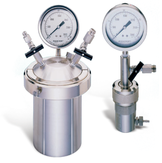
Parr stainless steel vessels for processing cell suspensions by the nitrogen decompression method are made in several sizes with full opening heads and self sealing closures which can be handled easily on any laboratory bench without special tools or fixtures. Each vessel has two valves and a pressure gage: one valve for charging with nitrogen and the other for withdrawing the homogenate and discharging it through an attached delivery tube. All of the fittings as well as the vessel itself are made of stainless steel with polished surfaces for good corrosion resistance and freedom from contamination. The individual parts are easily cleaned and can be thoroughly sterilised.
DESIGNS FOR LARGE AND SMALL SAMPLES
These vessels are made in five different sizes to accommodate samples ranging from 0.5ml to 5 litres, The maximum charging capacity for each vessel is limited to two-thirds of the internal volume of the vessel, but smaller samples can be treated in any of these vessels by simply placing the sample in a beaker or test tube and positioning it under the dip tube within the vessel.
The larger vessels, 4636, 4637 and 4638, are intended for large volume applications. Users are urged to run preliminary experiments in the 4635 vessel to confirm the suitability of the procedure before scaling up to these larger sizes.
EASY ACCESS TO VESSEL CHAMBER
Each of these vessles, except the small 45ml size, has a split-ring closure -an exclusive Parr design which allows the vessel to be opened or closed easily without disturbing any of the fittings or connecting lines attached to the head. In this closure two rings sections slide into place from the sides of the vessel to lock the head in position while a self-sealing O-ring maintains a tight seal at all pressures. The ring sections are secured by a steel retaining band that is raised from the bottom of the vessel and anchored with a single, hand-tightened screw.
On the small, 45ml vessel, a firm closure is obtained by simply turning down a knurled cap until it is hand tight. No wrenches or fixtures are required.
SAFETY PROTECTION
All Parr Cell Disruption Vessels are designed to accommodate the full 2000 psig available from a commercial nitrogen cylinder. If higher pressures should accidentally develop, the 920ml and larger vessels are protected by a safety rupture disc which will burst at approximately 3000 psig, well below the pressure at which any parts of the vessel would fail. A 3000 psi pressure gage and a 3000 psig rupture disc are standard but other gages and discs with lower pressure ratings can be furnished on special order.
INCLUDES NITROGEN FILLING CONNECTION
An 1831 nitrogen filling connection furnished with each vessel provides all of the fittings needed to fill the vessel from a commercial nitrogen cylinder. The 1823 connection consists of a control valve with a standard CGA-580 coupling for attachment to a nitrogen cylinder, a tank pressure gage and a flexible Nylon pressure hose for connection to the vessel inlet valve. Extra O-rings for the vessel head are included with each vessel.
CUSTOM MODIFICATIONS
Most cell disruption procedures can be handled readily in the 920ml, 4635 vessel with its customary fittings and attachments, yet applications may arise in which it will be desirable to modify a vessel to meet special requirements. It initial studies show a need for modifications, the user can return any vessel to the factory for such changes or additions as may be needed. Changes sometimes requested include:
- Multiple sample capacity. Up to three additional dip tubes and discharge valves can be installed in any vessel (except the 45ml size). With these additional outlets and appropriate sample holders, as many as four samples can be processed independently and simultaneously in a single unit.
- Special valves. Valves with either larger or smaller orifices can be provided. Users processing large quantities of materials that have a tendency to plug the orifice of the standard throttling valve may want to substitute either a larger needle valve or a ball valve to permit faster flow rates or to avoid stoppage. Fine metering valves with smaller orifices and very precise control capabilities may be helpful when treating samples that must be processed under very exact and repeatable conditions. Such valves may also be desirable when working with very small samples.
- Size conversion. Since cylinders for the 920ml and 1850ml vessels are interchangeable and both vessels use the same head and closure, any user of a 4635 vessel can increase the capacity of his unit to the 4636 size by simply purchasing a deeper cylinder (599HC2) and a longer diptube (591HC2). A similar conversion can also be made from the one gallon vessel to the two gallon size for users interested in vaccine production or in similar large volume applications. These larger vessels can also be equipped with vigorous internal agitators to speed equilibrium times when processing large volumes of material.
MAMMALIAN CELLS
Hunter and Commerford (1) published a paper in 1961 which has become a basic "cookbook" for the disruption of mammalian tissue by the nitrogen decompression method. Although most of the work reported by these authors was done with rat tissue, they also treated spleen, white cells, lymph nodes, tumors, thymus and other tissues to establish the general applicability of the method. Their results clearly demonstrated that cells can be disrupted by this method with minimum physical and chemical damage to the components.
H & C obtained complete disruption at pressures of 1300 psi and above, while pressures below 700 psi left whole cells and clumps of cells in the homogenate. At pressures between 800 and 1000 psi, cell-free homogenates were produced with nuclei intact. A hand press was used to pre-mince tissues prior to treatment in the vessel. The condition of the nuclei was found to be dependent upon the composition of the suspending buffer solution. Good results were obtained using isotonic solutions while nuclei swelling and rupture were observed in cells suspended in very dilute solutions. This was attributed to osmotic swelling which H & C found could be controlled by adding inorganic salts such as sodium chloride or organic solutes such as sucrose or glycerol. The nuclei were extremely fragile when the suspending medium contained no calcium, but the presence of as little as 0.0002M calcium chloride was found to stabilise the nuclei. Magnesium acetate is also useful for this purpose.
To determine the extent of damage to labile cells, H & C studied Deoxyribonucleoprotein, DNP, because of its susceptibility to chemical and physical stress, obtaining recoveries of over 90% DNP from the nuclear fraction with excellent preservation of the material. They also compared the enzyme activities of mitochondrial suspensions prepared by the nitrogen decompression method with suspensions produced in a PotterElvehjem homogeniser. No differences in enzyme activities were detected.
Dowben, Gaffey and Lynch (2) used the nitrogen decompression technique to prepare polyribosomes from L Cells, fibroblasts, human fetal cells from amniotic fluid, rat livers and muscle from chick embryos. Using 600 psi pressure they obtained better than 99.9% rupture and recovered more than 95% of the nuclei intact. Polysome yield was two to three times greater than when the cells were homogenised in a Dounce tissue grinder. In addition, they had better defined and more reproducible profiles. Significantly greater activities as measured by amino acid incorporation were also reported.
Short, Maines and Davis (4) compared the nitrogen decompression method with the Potter-Elvehjem types of PTFE pestle and glass tube homogenisers for preparing microsomal fractions for drug metabolism studies. The decompression method consistently produced over twice as much microsomal protein per gram of tissue as the pestle and tube fractionation. Enzyme activities per milligram of microsomal protein was found to be essentially the same for both methods, but it must be remembered that nitrogen decompression yielded over twice as much microsomal protein per gram of starting material.
Under microscopic examination the homogenates produced by the decompression method were found to be cell-free, while numerous cell clumps were observed in the pestle and tube homogenate. Electron microscopy of the microsomal pellets showed the particles to be smaller and more uniform in size for the decompression method. In summary, these authors stated that the nitrogen decompression method was more efficient and probably less variable than the PTFE pestle and glass tube methods.
Comparison with pestle and tube methods. In a recent application at the Veterans Administration Research Hospital in Chicago, a homogenate that had required eight hours to produce with the pestle and tube was prepared in fifteen minutes with a cell disruption vessel. In another laboratory, up to 12 kilograms of brain per day are being homogenised with a cell disruption vessel.
Wallach and his associates (5) have used the nitrogen decompression method to obtain complete cell fractionation with minimum nuclear damage. Working with Ehrlich Ascites Carcinoma Cells using a 0.0002M magnesium acetate buffer, they have studied the cellular distribution of phospholipides. Wallach has published many other papers in which the decompression technique has been used to prepare cell membranes.
VACCINE PREPARATION
A number of commercial laboratories have found that the nitrogen decompression technique is extremely effective for releasing virus from fertilised eggs. This method can be scaled up for commercial production using larger disruption vessels that are offered for this purpose by Parr.
BACTERIAL CELLS
Fraser (7) in 1951 published some of the earliest studies on nitrogen decompression and its effect on E Coli. Fraser's work was limited because his vessel was restricted to 900 psi operating pressure. Nevertheless, he was able to obtain 75% rupture in one pass and over 90% rupture in two successive passes using E Coli harvested during the log growth phase. Results with other bacteria and organisms with tough cell walls have been mixed.
There are several ways in which bacterial cells with tough walls can be treated to facilitate disruption by the nitrogen decompression method. These include: (1) harvesting the cells during an early growth phase before the full wall is developed; (2) growing the cells in the presence of an agent which will inhibit the formation of the cell wall; (3) using a lysozyme to weaken the wall prior to processing, or (4) using a mechanical pretreatment to weaken the cell walls before applying the nitrogen decompression method. Although these techniques have been applied successfully to many bacteria with heavy cell walls, they are not equally effective for yeast, fungus, spores and similar cells with very heavy or hard walls. Vigorous mechanical methods are generally required to break down the cell structure in these hard-walled materials since they generally do not respond well to treatment by the nitrogen decompression method.
PLANT CELLS
Loewus and Loewus (10) have published a number of papers in which they describe the application of nitrogen disruption procedures to plant cells and to tissue cultured plant cells. They also report considerable success in breaking diatoms by this method.
REFERENCES
(1) Hunter, M. J. and Commerford, S. L., 1961, "Pressure homogenization of mammalian tissues." Biochim. Biophys. Acta, 47:580- 6.
(2) Dowben, R. M., Gaffey, T. A. and Lynch, P. A., 1968. "Isolation of liver muscle polyribosomes in high yield after cell disruption by nitrogen cavitation." FEBS Letters, Vol. 2, No. 1, pages 1-3.
(3) Dowben, R. M., Lynch, P. M., Nadler, H. C. and Hsia, D. Y., 1969. "Polyribosomes from L. Cells." Exp. Cell Research, 58:167-9.
(4) Short, C. R., Maines, M. D. and Davis, L. E., 1972. "Preparation of hepatic microsomal fraction for drug metabolism studies by rapid decompression homogenization." Proc. Soc. Exper. Biol. Med., 140:58-65.
(5) Wallach, D. F. H., Soderberg, J. and Bricker, L., 1960. "The phospholipides of Ehrlich and ascites carcinoma cells composition and intracelfular distribution." Cancer Research, 20:397-402.
(6) Manson, L. A., Foshi, G. V. and Palm, J., 1963. "An association of transplantation antigens with microsomal pipoproteins of normal and malignant mouse tissues." J. Cell and ComD. Physiol., 61:109-18.
(7) Fraser, D., 1951. "Bursting bacteria by release of gas pressure." Nature, 167:33-4.
(8) Avis, P. J. G., 1967. "In subcellular components, preparation and fractionation." (Ed. Birnie, G. D. and Fox, S. M.) Chapt. 1, Pressure homogenization of mammalian cells. Published by Plenum Press, New York.
(9) Manson, L. A., 1972 "Extraction of membranous transplantation antigens by pressure homo- enization." (Ed. Kahan, B. D. and Reiifeld, R. A.) Chapt. 9,oyransplantation Antigens. Published by Academic Press, New York.
(10) Loewus, M. W. and Loewus, F., 1971. "The isolation and characterization of d-glucose 6-phosphate cycloaldolase (NDAdependent) from acer pseudoplatanus L. cell cultures. Plant Physiol. (1971) 48:255-260.